
Sorry this page is still under construction, will be available very soon.
Reconstructive Surgery
Scar Revision
Reconstructive Surgery
Removal of Skin Cancers
Sorry this page is still under construction, will be available very soon.
Reconstructive Surgery
Breast Reconstruction

Safety of Breast Reconstruction
It has been clearly documented that breast reconstruction do not affect the long-term survival of breast cancer patients. In other words, weather a breast reconstruction is done or not, this does not affect the outcome of the breast cancer.
On the other hand, it has also been clearly demonstrated that breast reconstruction has a profound positive effect on the psychological well being of patients. Patients feel better and is less phychologically affected by the mastectomy because the breast has been reconstructed. Patients can also go about their daily activity with less restrictions, such as putting on their clothes and going swimming.
Immediate Breast Reconstruction
The breast may be reconstructed immediately following removal of the breast cancer. With immediate breast reconstruction, the breast surgeon would employ a slightly different approach with the mastectomy, usually this would be what is called a SKIN SPARING MASTECTOMY. With the skin sparing sparing mastectomy technique, the skin of the breast (that is healthy and not affected by the cancer) is preserved. Preservation of the skin is advantageous because the natural breast skin gives a more natural shape to the breast when filled with either your own tissue or an implant.
Delayed Breast Reconstruction
This is when the mastectomy has been performed some time ago and patients would like to have her breast reconstructed. As the skin of the breast has been removed (usually leaving a straight scar), both additional skin and the breast would need to be reconstructed.
Techniques of Breast Reconstruction
The breast may be reconstructed with one of several methods. Your own tissues or implants may be used.
Breast Implants
Breast reconstruction using your own tissue (flap procedures)
The Latissimus Dorsi flap
The Latissimus Dorsi flap is a reliable and good flap for breast reconstruction, usually used with a breast implant. For this procedure the latissimus dorsi (a back muscle) with some overlying skin is brought to the front of the chest for the breast reconstruction. This provides added coverage over an implant and makes a more natural-looking breast than just an implant alone. This type of reconstruction can sometimes be used without an implant. To increase the fullness or volume of the flap to match the breast on the other side, fat grafting may be done at the time of the reconstruction.
Abdominal flaps (TRAM and DIEP)
Reconstructive Microsurgery
Sorry this page is still under construction, will be available very soon.
Reconstructive Surgery
Cleft Rhinoplasty
Sorry this page is still under construction, will be available very soon.
Reconstructive Surgery
Nose Fractures
Nasal fracture is the commonest fracture of the facial bones. Fracture can occur following low velocity injury like a fall or following a high velocity injury such as a road traffic accident. It may occur in isolation or may be associated with other fractures of the face such as the cheekbone. With nose fractures, generally there may be some bleeding from either or both nostrils. The bleeding generally stops by itself. The patient may notice that the nose is crooked, swollen or bruised. Breathing may be impaired as well as the fractures may have obstructed one or both of the nasal passages. Because of the injury and swelling, the degree or severity of the nasal deviation is generally difficult to appreciate in the acute/early stage.

Figure 2: A saddle nose as a result of nasal fracture.
Reconstructive Surgery
TREATMENT
Generally it is recommended that the fractured bones be precisely repositioned by manipulation under general anesthesia. This procedure is known as M&R of the nose (Manipulation and Reduction of the nose). The nose is then packed and splinted externally by a splint to prevent the repositioned bone pieces from moving.
The splints and packs are progressively taken out over 1 week and the patient needs to protect the nose from pressure and movements over the next 3 weeks. The patient needs to be followed up long term. With this approach however, a significant proportion of patients may have a residual crooked nose. A definitive open rhinoplasty for crooked nose may be performed from 4 – 6 months after the initial injury.
IMMEDIATE RHINOPLASTY FOLLOWING NASAL FRACTURES
Tummy Tuck
Abdominoplasty
Tummy tuck surgery, also known as abdominoplasty, removes excess fat and hanging skin from the lower abdomen. In most cases, the separated rectus abdominis muscles (known as diastasis of the recti) is also repaired at the same time. This restores the abdominal musculature that has weakened as a result of pregnancies thereby improving the shape as well as the function of the abdomen.

Why do a Tummy Tuck?
Tummy tuck is an effective procedure to correct a lower abdomen pannus that protrudes or is loose and sagging. The most common causes of this include: aging, pregnancy, prior surgery and significant weight gain or loss In many patients, particularly in female patients with previous pregnancies, the abdominal muscle (rectus abdominis) separates (called diastasis of recti), resulting in an area of weakness in the central abdomen that bulges when the patient strains or exerts. This bulge is a type of hernia (called ventral hernia) and may be associated with symptoms such as pain in the central abdomen or back pain, poor posture, pelvic floor problems and bloating. The diastasis of the recti may be repaired during the tummy tuck, effectively relieving these symptoms.
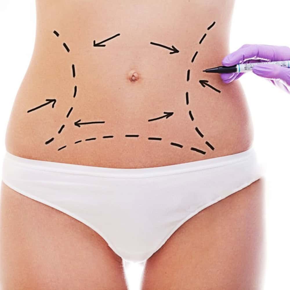
How is the tummy tuck done?

Recovery from a Tummy Tuck
You will need to take at least two weeks off from work for the recovery following a tummy tuck surgery. While you recover, you’ll need to stop physical activity for about one month after surgery. Mild pain may be experienced and would be adequately treated with oral painkiller to for the first few days after surgery. You should also avoid heavy lifting for 2 months after the surgery.
Gynaecomastia Treatment
Male Breast Reduction
Male breast development, known scientifically as Gynaecomastia, is defined as excessive development or growth of the male breast. This condition affects up to 36% of males and usually affects both breasts. The cause of the male breast enlargement is usually a hormonal imbalance, with relative reduction of male hormones (androgens) and relative excess of female hormones (estrogens). In majority of patients, the cause of this imbalance remains unknown, with most patients having a normal hormonal profile.
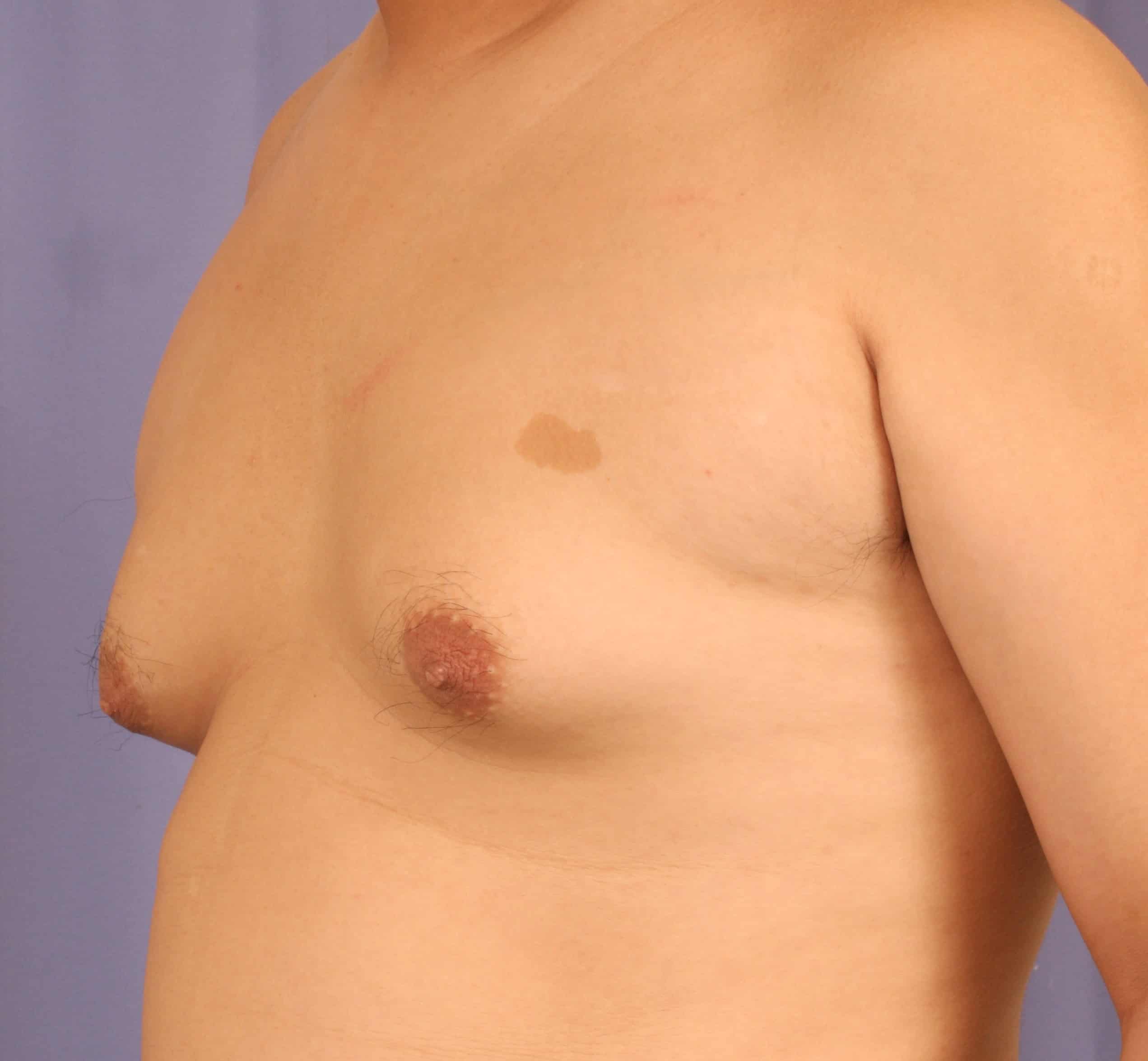
Natural History of Gynaecomastia
Many patients are affected by gynaecomastia during puberty, and in majority of patients, the condition usually spontaneously regress over 3 to 18 months. Weight loss may help in some patients.
Patients with gynaecomastia for longer than this duration and into early adulthood rarely regress and is an indication for surgical removal.
It has been proven that adolescent gynaecomastia has a significant negative impact on the psychological well-being of affected patients, specifically with regards to social functioning, mental health and self-esteem. Of note, the negative impact was not affected by the severity or grade of the gynaecomastia. Early treatment is therefore recommended regardless of the severity of the gynaecomastia in adolescent patients.
Indications for surgery
Patients may consider surgical treatment for the gynaecomastia if:
- The condition has been present for more than 12 – 18 months without regression.
- It is causing the patient psychological stress and affecting social activities
- If it is impeding his daily activities like exercises
Physical Examination
The breast is examined to determine the tissue characteristics of the individual patient’s enlarged breast. The nipple disc is palpated to determine the extent of enlargement of the breast tissues. This is generally firmer and harder than the surrounding tissues, which is primarily fat and therefore much softer.
Surgical Techniques
Depending on the severity of the breast enlargement, tissue characteristic of the individual patient’s breast as well the extent of skin excess (drooping) of the breast, one of several techniques may be used alone or in combination.
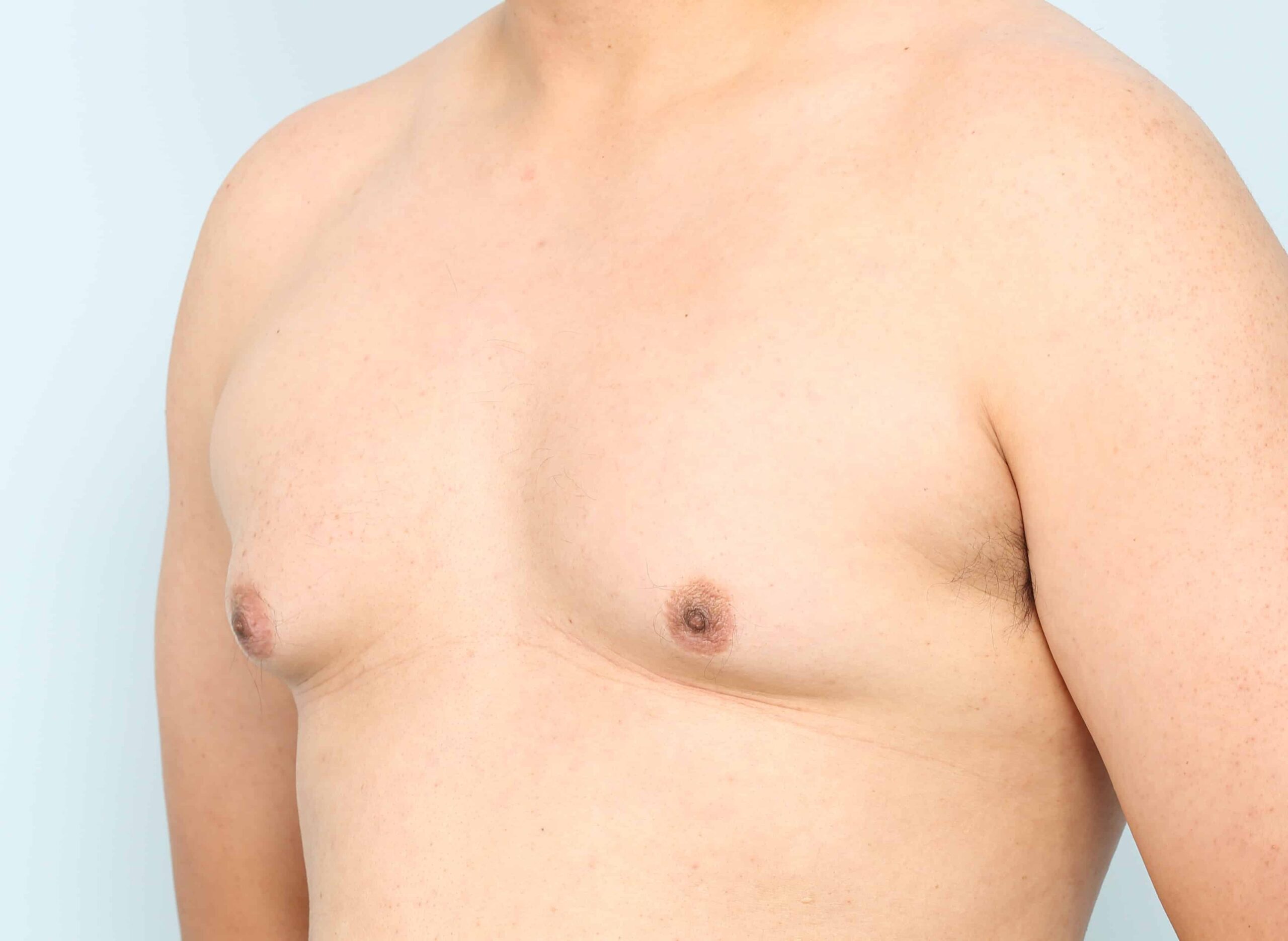
Minimally Invasive Techniques
LIPOSUCTION & MICRODEBRIDER technique
Liposuction is used to remove the fatty tissue around the enlarged breast. Power assisted liposuction or Ultra sound assisted (VASER) liposuction may be used with good effect. The use of the microdebrider is primarily to enable more effective and complete removal of the firm breast disc behind the nipple. While liposuction alone does remove the disc to some extent, I have found the combine use of liposuction and microdebrider give a more even and predictable reduction/ flattening of the male breast. This combination of techniques is the most commonly used approach today as it is effective while being done with minimal incisions (2 stab incisions only per chest) thereby minimizing the scarring associated with the procedure.
Open Techniques
Breast Lift
This is for patients with more severe forms of breast enlargement usually seen in massive weight loss patients where the degree of sagging is beyond what contraction of the skin after the liposuction can predictably correct. In these patients, a skin tightening procedure call a breast lift is needed to remove the excess skin. Incisions or cuts will be needed to remove the excess skin and to flatten the chest and the placement of these incisions will be discussed with the patients.
Post Operative care
After surgery, to promote more rapid wound healing and contraction of the skin, a course of radiofrequency treatment to the treated areas would be prescribed. Patients will be encouraged to wear a firm compressive pressure garments for up to 2 months after the procedure and be taught to perform firm massages on the area.
In Summary
Male breast reduction is a predictable and safe procedure. It had profound physchological as well as aesthetic benefits for patients affected for this condition. To discuss more in detail about your specific needs as well as review some before and after photos of our patients, please come in for a consultation.
Facial Aesthetic Plastic Surgical Procedures
Eye Bags & Lower Eyelids
The eyes are one of the first things people notice when looking at someone, so it is no surprise that eye bags are one of the most common complaints when a patient visits a plastic surgeon. As eye bags tend to make the patient look tired and listless, removal of the ‘bags’ can rejuvenate the patient’s face by making them look fresh and younger.
In general, patients with eyes bags can be divided into two groups with different severity of midcheek aging, and each requiring different surgical approaches:
- Patients with true excess of eye bag fats, with no other signs of midcheek aging: These patients are generally below 40 years old.
- Patients with eye bags and associated with midcheek aging: These are generally patients above 40 years old.
These 2 groups of patients will be taken into consideration:
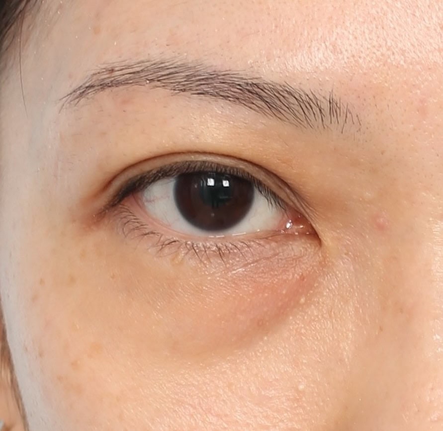
Young patients with eyebags only
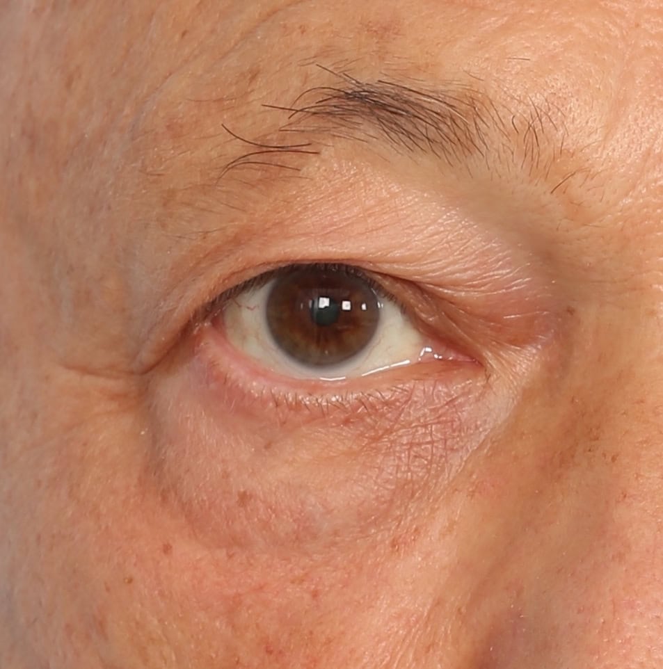
Rethinking Eye Bags
Patients with eyebags with significant midcheek aging
Rethinking eyebag surgery: Looking beyond the eyebags to achieving TRUE midcheek rejuvenation
Eye bags are one of the most common cosmetic concerns patients tend to have. As patients request for their eye bags to be removed, excision of the orbital fat pads that is responsible for the ‘bags’ has been the most common procedure for many years. While the fat excision or removal approach is still valuable and valid in selected patients, it may not be the best solution for many others with eye bag problems.
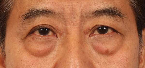
Figure 1
Eyebags have the visual effect of making a person look tired
First, lets consider what the patients are really seeking beyond correction of the eyebags. While the eyebags are quite noticeable, when we look deeper, it is ‘tired look’ that has gradually replaced the ‘fresh’ look that the patients once possess in their youth that so burden the patients. One that makes them appear to lack vitality even though they maybe full of energy inside. This appearance can be attributed to the complex changes of the midcheek that occur with aging.
A fresher, more vibrant looking you.
Let’s take a look at the deeper concerns of patients with eye bag problems, recent advances in the understanding of the anatomy of the aging midcheek (of which the eye bags is part of) and the elements needed to achieve optimal midcheek rejuvenation.
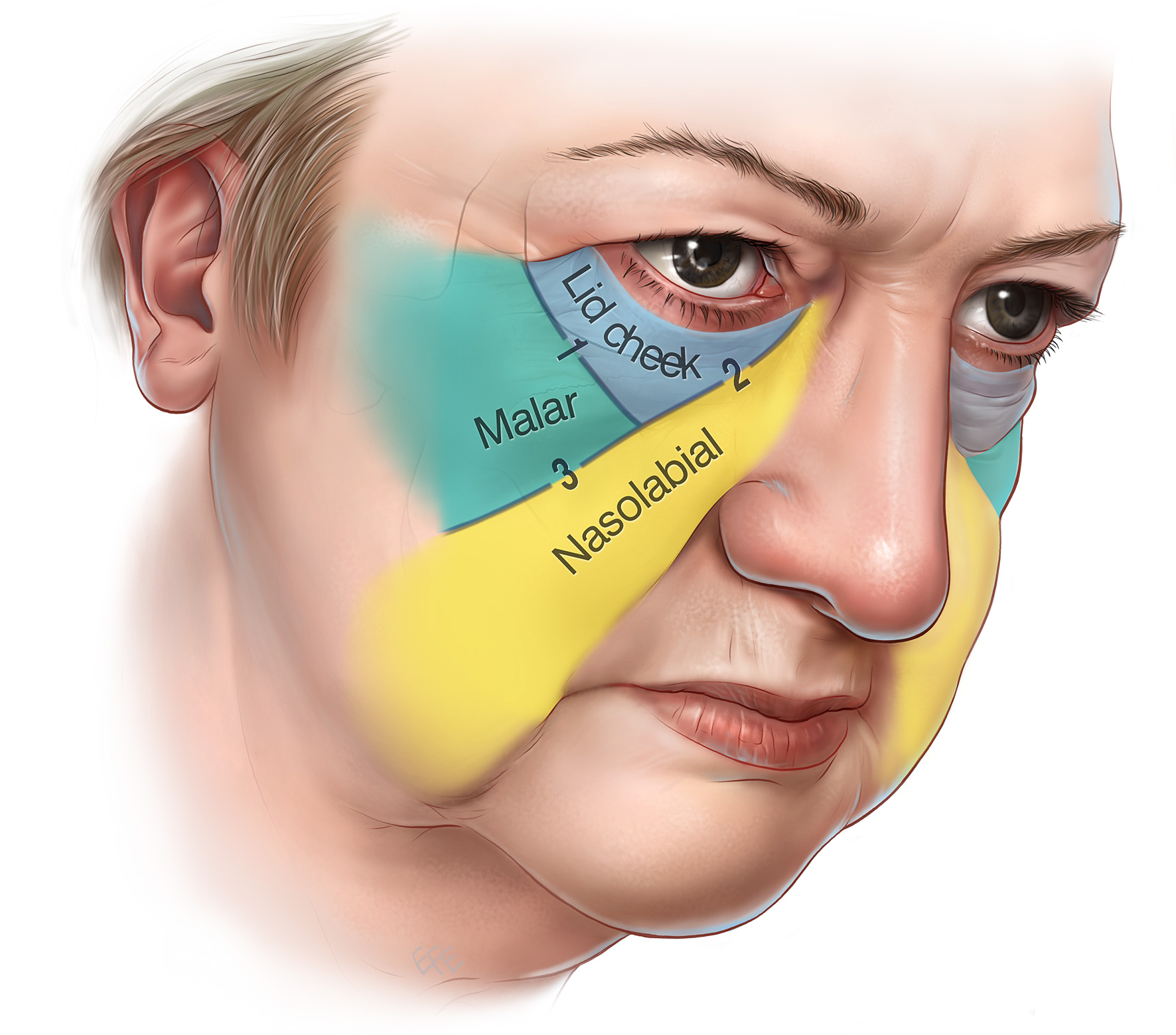
Figure 2
Segmentation of the midcheek into 3 segments with the aging midcheek
First, lets consider what the patients are really seeking beyond correction of the eye bags. While the eye bags are quite noticeable, often what bothers the patients most is losing the ‘fresh look’ that they once had in their youth Looking ‘tired’ makes them appear to lack vitality even though they maybe full of energy inside. This can be attributed to the complex changes of the midcheek as we age.
At a deeper level, these patients are actually seeking rejuvenation of the midcheek for a fresh and youthful look. Most importantly, the eye bags are only one aspect of the aging midcheek, and addressing the eye bags alone is inherently limited by what the approach can achieve. As such, all aspects of the aging midcheek have to be addressed to achieve the highest level of rejuvenation.
The anatomy of the midcheek
The aging midcheek comprises several components, one of which is the bulging of the orbital fat that is commonly known as eye bags. This is also perhaps the most noticeable change that comes with aging.
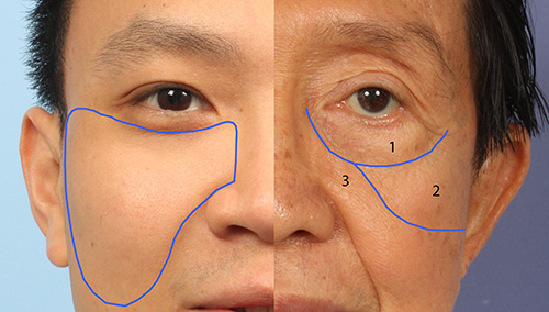
Figure 3
midcheek
In youth the midcheek is full and rounded with no eyebags (left side). With aging, the midcheek segments into 3 segments with deflation and sagging clearly seen (right side).
Equally important is the portion of the cheek, immediately below the eye bag. The soft tissues here tend to deflate and descend with aging, and this is particularly obvious in the inner aspect of the midcheek. Another significant change is the retrusion or resorption of the midface skeleton. This contributes profoundly to the loss of soft tissue support for the aging midcheek. Finally the skin thinning and fixed wrinkling all also play a role in the appearance of the aging midcheek. In particular, deflation is an aspect of the aging midcheek that is increasingly recognised as an important contributory factor. One only has to observe the fullness in the midcheek that is so evident in youth to appreciate the importance of volume restoration in midcheek rejuvenation.
A recent anatomical advance is the understanding of the facial retaining ligaments of the midcheek.
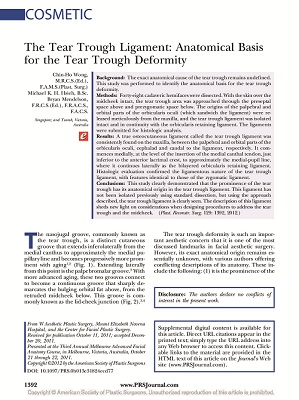
Article
The tear trough-orbicularis retaining ligament complex
In this article, Dr Wong and colleagues described the tear trough-orbicularis retaining ligament complex, the key structure that holds the midcheek soft tissues. Utilizing this anatomy, midcheek lift can effectively and safely be done.
The retaining ligaments are discrete fibrous structures the bind the tissue layers together. It is because of its function that it manifest as the surface grooves we see with aging. A prime example of this is the tear trough below the eye bags. Precise release of these ligaments can effectively soften these grooves, playing an integral part in rejuvenation of the midcheek.
By understanding the factors that contribute to the tired look, we can then design procedures to address this issue and the components of the aging midcheek.
When it comes to conventional eye bag surgery, excision is often the prescribed solution for patients with eye bags. While fat excision alone is effective in selected patients, especially patients with ‘true’ excess of the orbital fat, fat removal alone is inherently limited by what the procedure can achieve in patients with more advanced midcheek aging. When carried out well, removal of the fat would eliminate the eye bags. However, quite often this does not produce the refreshed look that patients are seeking.
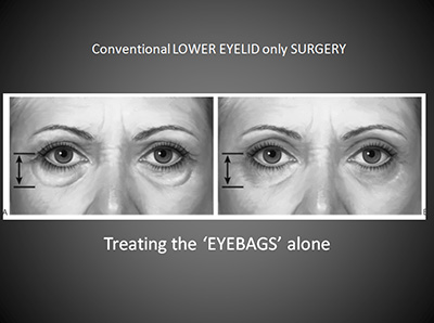
Figure 4
advanced midcheek aging
Removal of eyebag alone in patients with more advanced midcheek aging have the effect of hollowing out the orbits. This does not rejuvenate the midcheek but makes the person “old” from the hollowed out appearance.
Due to the significant deflation, loss of volume as well as the descent in tissue, all these aspects have to be addressed to achieve a refreshed look. Accordingly, in rethinking our approach to the common complaint of eye bags, and depending on the patients’ specific needs, the following may warrant consideration:
- The eye bags: This may be removed in cases of true excess or more commonly, the fat may need to be preserved but REDISTRIBUTED to move it from area where it is not wanted to that part of the cheek where VOLUME is deficient.
- For the descended cheek tissues, a CHEEK LIFT is needed to reposition the cheek mass into a more YOUTHFUL position
- For patients with more significant loss of volume or deflation of the midcheek, these need to be restored. This can be achieved either by synthetic fillers (such as hyaluronic acids), cheek augmentation with implants or micro fat grafting, which is becoming increasingly popular.
In rethinking eyebags therefore, the following can be said
- In patients with true orbital fat excess, the fat excision technique continue to be an effective approach when it comes to its correction
- In patients with significant aging changes wanting to achieve a fresh and rejuvenated appearance, a more advanced approach incorporating fat redistribution, midcheek lift and midcheek augmentation with fat grafting is currently the most powerful technique available.
Please visit us for an in-depth discussion on your specific needs.
The eyes are the mirror of the soul - Proverb
Asian Upper Eyelids
The upper eyelid crease in Asians makes the eyes appear larger and the person more friendly and approachableOur personality and attitudes are expressed through our eyes. The upper eyelids have a profound impact on the appearance of the eyes. It determines if you look young and fresh or tired or even fierce. A well-placed and crisp upper eyelid crease frames the eyes, focusing the observer on the eyes and, in so doing, brings the sparkle out of the eyes. An absent upper eyelid crease (that occurs congenitally or as a result of hooding that occurs with aging), in contrast, shifts the focus of the observer to the upper eyelid, giving the visual impression that the eyes are smaller and the upper eyelids as overhanging the eyes. This makes the person appears reclusive, listless and distant.
Upper eyelid surgery is for 2 groups of patients: For patients with an absent or poorly formed upper eyelid fold and for patients with significant aging of the upper eyelids.

Excessive skin and fat excision with traditional techniques results in sunken eyes and upper eyelid hollowing (above). With aging, the skeleton becomes visible giving an emanciated appearance
ABSENT or ASYMETRIC UPPER EYELID CREASE

An Asian patient with an absent upper eyelid crease with no excessive upper eyelid fat or puffiness
With this in mind, upper eyelid crease creation can be done by 2 methods in Asians.
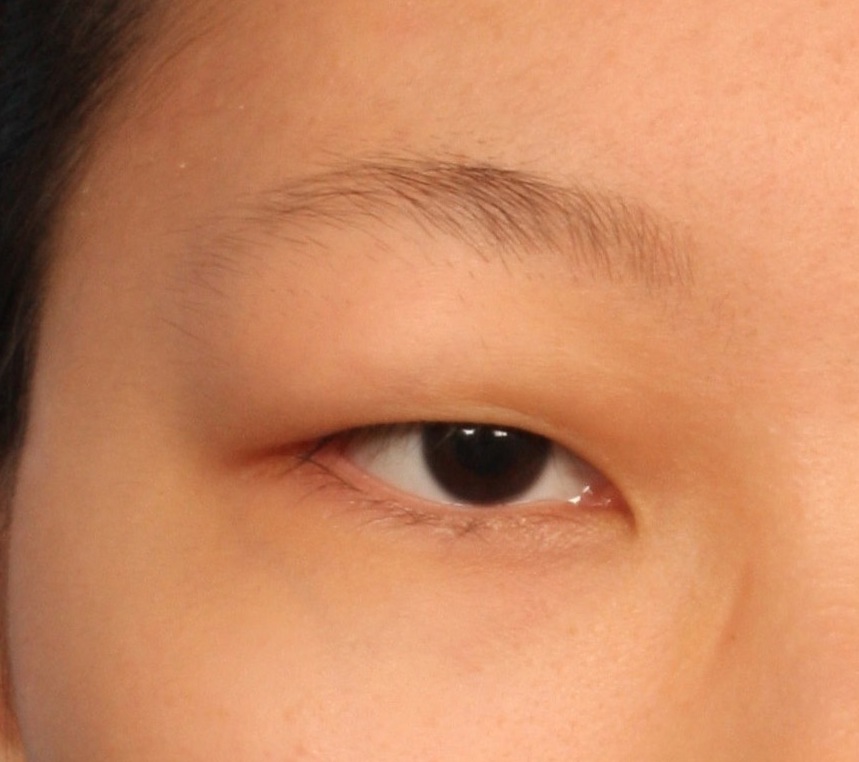
Patients with puffy upper eyelids and a true excess of upper eyelid fats would benefit from the incision technique as it allows for precise removal of upper eyelid skin excess, muscle as well as orbital fat
INCISION UPPER BLEPHAROPLASTY FOR CREATION OF DOUBLE EYELID CREASE IN YOUNG PATIENTS
Incision upper blepharoplasty is recommended for younger patients who are seeking to create natural and crisp upper eyelid crease to brighten the eyes. The incision technique has the advantage of allowing for more predictable correction of minor asymmetries of the upper eyelid, removal of true excess upper eyelid fat as well as for the formation of a permanent upper eyelid crease with minimal chance of loosing the fold. The versatility afforded by the incision technique is that the amount of skin and muscle that is excised can be precisely controlled in designing the procedure and that the shape and contour of the fold that best suit the patient can be designed with the patient’s input. Dr. Wong has published his technique of incision upper blepharoplasty with the Hinge Upper Blepharoplasty technique. You may review this paper here.
As this technique involves small cuts of the upper eyelids, swelling is a little more compared with the suture technique, lasting about 1 – 2 weeks.
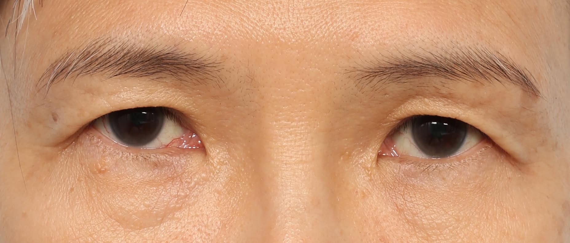
INCISION UPPER EYELID SURGERY FOR THE AGING UPPER EYELIDS